
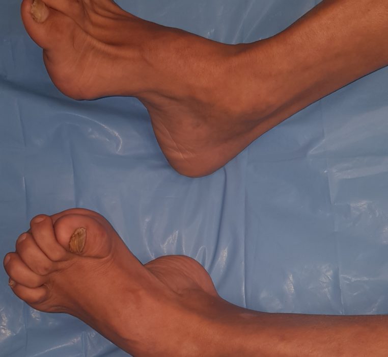
CAVUS FOOT / High Medial Arch foot deformity RECONSTRUCTION
High medial arch foot or pes cavus foot abnormality is a condition causing one to place too much weight and stress on the ball and heel of the foot in erect posture while standing and/or walking.
It is also commonly associated with other foot and ankle deformities like clawing of the toes, posterior hind foot deformity, contracture of the plantar fascia, equinovarus, calcaniovarus and cock-up deformity of the great toe. As a result of pes cavus deformity, one can develop metatarsalgia and calluses because of increased weight bearing over the metatarsal heads.
Types of Pes cavus
Types of Pes cavus
Three types based on aetiology, clinical signs and radiological appearance:
- Pes cavovarus, the most common. Radiologically the forefoot is plantarflexed in relation to the rearfoot.
- Pes calcaneocavus , is mainly seen following paralysis of the triceps surae due to poliomyelitis, the calcaneus is dorsiflexed and the forefoot is plantarflexed. Radiologically there is a large talo-calcaneal angle.
- Pes cavus, the calcaneus is highly arched and is neither dorsiflexed nor in varus position. Forefoot is in plantarflexed position on the rearfoot.
Types based on location of APEX of the deformity
- Anterior Cavus (Forefoot Cavus)
–Local
–Global
- Metatarsus cavus
- Posterior Cavus
- Combined
Clinical Picture
- Lateral foot pain from increased weight bearing on the lateral foot,
- instability,
- difficulty walking and problems with footwear
- metatarsalgia,
- pain under the first metatarsal,
- plantar fasciitis,
- painful callosities,
- ankle arthritis,
- achilles tendonitis
- keratosis
- lateral ankle instability
- hindfoot varus
- The forefoot plantar flexion
- hindfoot varus
- lower limb stress fractures
- knee pain
- iliotibial band friction syndrome
- back pain
- tripping
Medical management
At Pakistan Orthopaedic and Cosmetology Center, our aim of medical management is to allow the patient to ambulate without any problems. It is important for the patient to understand that surgical reconstruction does not provide a normal foot. The main goal of surgical reconstruction is to produce a plantigrade foot and pain relief. Repeated surgical procedures can be necessary, especially if the deformity is progressive. Surgical procedures can be broadly categorized into soft-tissue and bony procedures. Tendon transfers and osteotomies can provide correction of the deformity without requiring an arthrodesis.
Surgical Management
- Correcting a cavovarus foot
- Most of the corrections involve tendon transfers and capsular and facial releases
- Correction of plantar flexion of the first ray by performing a dorsiflexion
- ST tarso-metatarsal arthrodesis.
- Reduction of hindfoot varus by performing a lateralizing calcaneal osteotomy.
- Arthrodesis 1st TMT joint, lateral calcaneal osteotomy for hind foot.
For further details:
Subscribe and watch: our Youtube channel
Visit lso: Facebook page
call us on WhatsApp:
+923015943329
+923339113796